Know Infertitlity
Infertility treatments
Diagnosing InfertilityMale:
Obstetrics and Gynaecology
Infertility Treatment Services
Andrology |
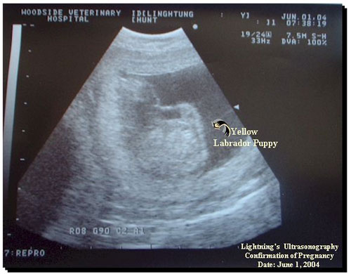
UltrasonographyUltrasonography is a widely used ultrasound based diagnostic imaging tool used to view internal organs, muscles, bones and determinetheir size and structure and in turn diagnose a wide variety of diseases and conditions with real time tomographic images. In ultrasonography, high-frequency sound waves are bounced off through body tissues and the resulting echoes are recorded and transformed into graphic images. This technique is also used to view fetuses during routine and emergency prenatal care. Such applications during pregnancy are referred to as obstetric sonography. Obstetric SonographyObstetric ultrasound technique is used routinely in obstetric appointments during pregnancy to detect conditions that would pose danger to the mother and the baby. Routine ultrasound in early pregnancy (less than 24 weeks) appears to enable early detection of multiple pregnancies, better gestational age assessment and earlier detection of clinically unsuspected fetal malformation at a time when termination of pregnancy is possible.Obstetric ultrasound is primarily used to:
 Ultrasonography procedureThis technique makes use of an abdominal or vaginal probe or transducer depending on the stage of the pregnancy. The probe emits high frequency sound waves into the body which are allowed to bounce off different organs and are sent back as electrical signals. These signals are then processed and displayed as images or graphs. Vaginal ultrasound is done earlier on in pregnancy and is also referred to as transvaginal ultrasound. When is the test doneThis test can be done at any stage of pregnancy although routine ultrasound in early pregnancy (less than 24 weeks) allows the determination of multiple pregnancies and earlier detection of fetal malformation at a time when termination of pregnancy is possible. AdvantagesUltrasound technology is portable and relatively inexpensive, especially in comparison to other techniques such as magnetic resonance imaging (MRI) and computed tomography (CT). Other advantages include:
Risks involvedThe long term effects of repeated ultrasound exposures on the fetus are not fully known. It is advised that ultrasound only be used if medically indicated.
|
Our TeamNews & EventsClinic LocationVideo Gallery |


